Diagram Of The Muscles In The Forearm - Anatomy Human Forearm Muscles Wall Decal Design 1 Wallmonkeys Com : From the arm muscle diagram above, the muscles of the arm that can be seen easily on the surface include biceps, triceps, brachioradialis, extensor carpi radialis longus, and deltoid.
Diagram Of The Muscles In The Forearm - Anatomy Human Forearm Muscles Wall Decal Design 1 Wallmonkeys Com : From the arm muscle diagram above, the muscles of the arm that can be seen easily on the surface include biceps, triceps, brachioradialis, extensor carpi radialis longus, and deltoid.. The superficial extensors of the forearm are the brachioradialis, extensor carpi radialis longus, anconeus, extensor carpi radialis brevis, extensor carpi ulnaris, extensor digitorum and extensor digiti minimi. The muscles in the forearms are numerous and complex. To begin, spend some time looking at the forearm muscles diagram above. They control movements of the wrist, hand, fingers and thumb. Write down the muscles of the forearm selected by your instructor and, for each, give the location of that muscle and what effect contracting that muscle has.
When the biceps contracts, it pulls the forearm up and rotates it outward. We are pleased to provide you with the picture named right arm muscle and tendon anatomy.we hope this picture right arm muscle and tendon anatomy can help you study and research. To begin, spend some time looking at the forearm muscles diagram above. Muscles of upper back 12 photos of the muscles of upper back map of upper back muscles, muscles of the upper back and chest, origin and insertion of upper back muscles, superficial muscles of the upper back, tight muscles of the upper back and neck, human muscles, map of upper back muscles, muscles of the … The superficial extensors of the forearm are the brachioradialis, extensor carpi radialis longus, anconeus, extensor carpi radialis brevis, extensor carpi ulnaris, extensor digitorum and extensor digiti minimi.
![]()
For the most part, they are polyarticular.
An extensor muscle of the elbow joint and an antagonist of the biceps and brachialis muscles. Biceps are large muscle of the upper arm is formally known as the biceps brachii muscle, and rests on top of the humerus bone. Here you can see all the extensor forearm muscles clearly labeled. Rate 5 stars rate 4 stars rate 3 stars rate 2 stars rate 1 star. We are pleased to provide you with the picture named different types of muscles of arm diagram.we hope this picture different types of muscles of arm diagram can help you study and research. Learn vocabulary, terms, and more with flashcards, games, and other study tools. Rotates the forearm so the palm is facing the ceiling). It is attached, behind, to the olecranon and dorsal border. The arm is one of the body's most complex and frequently used structures. Anatomy of the human body. The muscles of this chapter are involved with motions of the forearm (radius and ulna) at the radioulnar joints, the hand at the wrist (radiocarpal) joint, and the fingers at the metacarpophalangeal (mcp) and/or the proximal interphalangeal (pip) and distal interphalangeal (dip) joints; The superficial extensors of the forearm are the brachioradialis, extensor carpi radialis longus, anconeus, extensor carpi radialis brevis, extensor carpi ulnaris, extensor digitorum and extensor digiti minimi. Overview diagram showing the labeled forearm extensor muscles forearm muscles (extensors) labeled and unlabeled.
Anatomy of the human body. Overview diagram showing the labeled forearm extensor muscles forearm muscles (extensors) labeled and unlabeled. The superficial extensors of the forearm are the brachioradialis, extensor carpi radialis longus, anconeus, extensor carpi radialis brevis, extensor carpi ulnaris, extensor digitorum and extensor digiti minimi. Deep fascia of the forearm).—the antibrachial fascia continuous above with the brachial fascia, is a dense, membranous investment, which forms a general sheath for the muscles in this region; The muscles and fasciæ of the forearm antibrachial fascia (fascia antibrachii;

These types of strains are quite severe and involve complete rupture of the muscle fibers and tendons.
Flexor carpi ulnaris, palmaris longus, flexor carpi radialis, and pronator teres. It is called lister's tubercle. It can also fixate the elbow joint when the forearm and hand are used for fine movements, e.g., when writing. Forearm muscles anatomy the term forearm is used in anatomy to distinguish it from the arm, a word which is most often used to describe the entire appendage of the upper limb, but which in anatomy, technically, means only the region of the upper arm, whereas the lower 'arm' is called the forearm. It rotates the forearm and also flexes the elbow. Superficial posterior muscles of the forearm posterior compartment muscles of the forearm. Rate 5 stars rate 4 stars rate 3 stars rate 2 stars rate 1 star. Brachioradialis is one of the muscles that comprise the posterior compartment of the forearm. The muscles and fasciæ of the forearm antibrachial fascia (fascia antibrachii; The extrinsic hand muscles originate in the forearm and insert on structures within the hand. For more anatomy content please follow us and visit our website: Grade iii strain of forearm muscle: The large muscle of the upper arm is formally known as the biceps brachii muscle, and rests on top of the humerus bone.
The tendon then attaches to the most distal bone in the thumb. The arm is one of the body's most complex and frequently used structures. (the lower arm is the forearm or antebrachium.) there are three muscles on the upper arm that are parallel to the long axis of the humerus, the biceps brachii, the brachialis, and the triceps brachii. Related posts of muscles of the arm and forearm diagram muscles of upper back. We are pleased to provide you with the picture named right arm muscle and tendon anatomy.we hope this picture right arm muscle and tendon anatomy can help you study and research.
The muscles that extend the hand at the wrist are located on the posterior portion of the forearm.
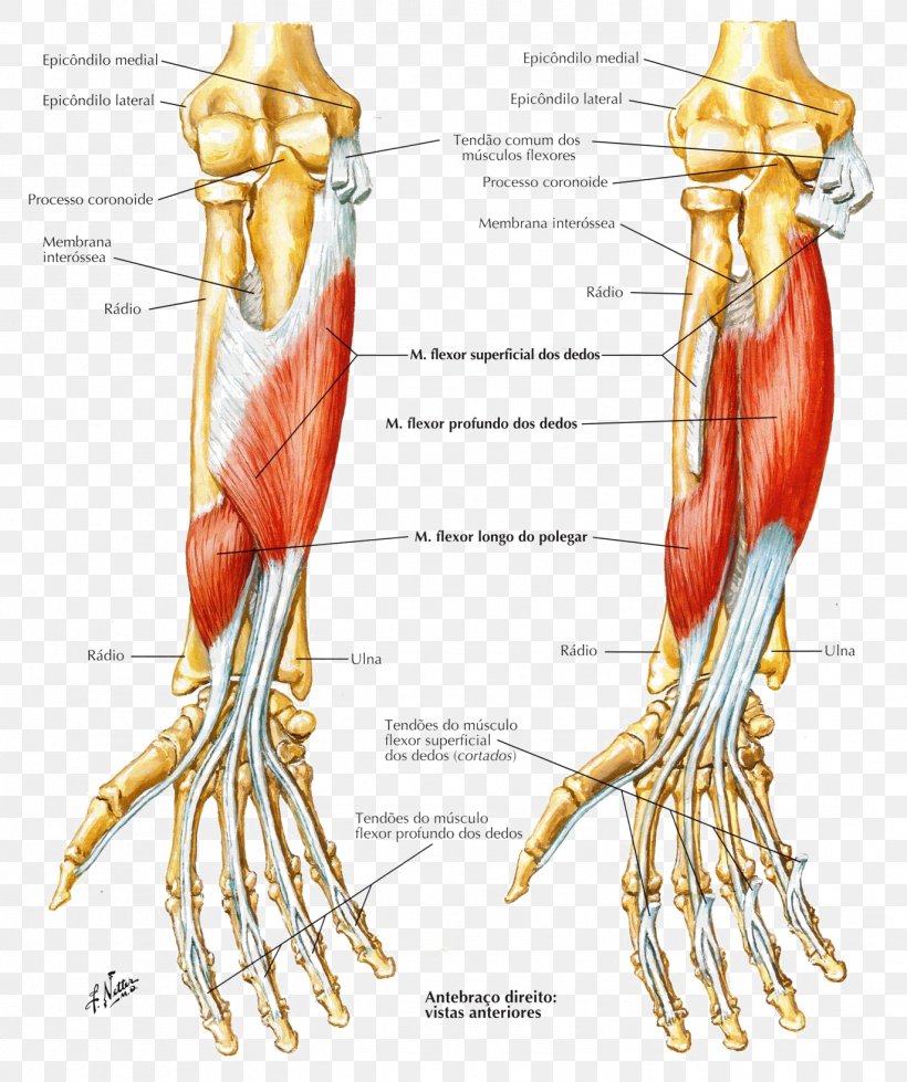
From the arm muscle diagram above, the muscles of the arm that can be seen easily on the surface include biceps, triceps, brachioradialis, extensor carpi radialis longus, and deltoid. It is the most superficial muscle of the radial side of the forearm, forming the lateral wall of the cubital fossa. Once you're ready, you can try labeling the muscles for yourself using the blank forearm. Here you can see all the extensor forearm muscles clearly labeled. An upper arm muscle composed of 2 parts, a long head and a short head. Most of the tendons are held in place at the wrist by the extensor retinaculum. Rate 5 stars rate 4 stars rate 3 stars rate 2 stars rate 1 star. Head muscles forearm muscles bones and muscles anatomy organs human body anatomy human anatomy and physiology muscle diagram body diagram diagram chart. We are pleased to provide you with the picture named right arm muscle and tendon anatomy.we hope this picture right arm muscle and tendon anatomy can help you study and research. Diagram of the forearm flexors. An extensor muscle of the elbow joint and an antagonist of the biceps and brachialis muscles. The muscles in the forearms are numerous and complex. Such forearm muscle strains may result in mild loss of strength of the forearm muscles.


Komentar
Posting Komentar